
Diabetic Retinopathy
Diabetes can affect sight
If you have diabetes mellitus, your body does not use and store sugar properly. High blood-sugar levels can damage blood vessels in the retina, the nerve layer at the back of the eye that senses light and helps to send images to the brain. The damage to retinal vessels is referred to as diabetic retinopathy.
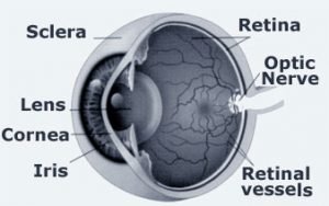 Normal Eye
Types of diabetic retinopathy
There are two types of diabetic retinopathy:
nonproliferative diabetic retinopathy (NPDR)
proliferative diabetic retinopathy (PDR).
NPDR, commonly known as background retinopathy, is an early stage of diabetic retinopathy. In this stage, tiny blood vessels within the retina leak blood or fluid. The leaking fluid causes the retina to swell or form deposits called exudates.
Many people with diabetes have mild NPDR, which usually does not affect their vision. When vision is affected it is the result of macular edema and/or macular ischemia.
Macular edema is swelling, or thickening, of the macular, a small area in the center of the retina that allows us to see fine details clearly. The swelling is caused by fluid leaking from retinal blood vessels. It is the most common cause of visual loss in diabetes. vision may be mild to severe, but even in the worst cases, peripheral vision continues to function.
Macular ischemia occurs when small blood vessels close. Vision blurs because the macular no longer receives sufficient blood supply to work properly.
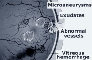 Common problems associated with diabetic retinopathy
PDR is present when abnormal new vessels begin growing on the surface of the retina or optic nerve. The main cause of PDR widespread closure of retinal blood vessels and, preventing adequate blood flow. The retina responds by growing new blood vessels in an attempt to supply blood to the area where the original blood vessels closed.
Unfortunately, the new, abnormal blood vessels do not re-supply the retina with normal blood flow. The new vessels are often accompanied by scar tissue that may cause wrinkling or detachment of the retina.
PDR may cause more severe vision loss than NPDR because it can affect both Central and peripheral vision.
Proliferative diabetic retinopathy causes visual loss in the following ways:
Vitreous hemorrhage: the fragile new vessels may bleed into the vitreous, a clear jelly-like substance that fills the center of the eye. If the vitreous hemorrhage is small, a person might see only a few new dark floaters. A very large hemorrhage might block out all vision.
It may take days, months or even years to resorb the blood, depending on the mountain blood present. If I does not clear vitreous fluid adequately within a reasonable time, vitrectomy surgery may be recommended.
The vitreous hemorrhage alone does not cause permanent vision loss. When the blood clears, visual activity may return to its former level unless the macula is damaged.
Traction retinal detachment: when PDR is present, scar tissue associated with neovascularization can shrink, wrinkling and pulling the retina from its normal position. The macular wrinkling can cause visual distortion. The more severe vision loss can occur if the macula or large areas of the retina are detached.
Neovascular glaucoma: occasionally, extensive retinal vessel closure will cause you, abnormal blood vessels to grow on the iris and block the normal low of fluid out the eye. Pressure in the eye builds up, resulting in neovascular glaucoma, a severe eye disease that causes damage to the optic nerve.
How is diabetic retinopathy diagnosed?
A medical eye examination is the only way to find changes inside your eye. An ophthalmologist can often diagnose and treat serious retinopathy before you are aware of any vision problems. The ophthalmologist dilates your pupil and looks inside of the eye with an ophthalmoscope.
If your ophthalmologist finds diabetic retinopathy, he or she may order color photographs of the retina or special test called fluorescein angiography to find out you need treatment. In this test a dye is injected in your arm and photos of your eye are taken to detect where fluid is leaking. Your Ophthalmologist may also order a special test called an OCT. This test is able to image the retina, measure the thickness of the retina and find areas of swelling within the retina.
How is diabetic retinopathy treated?
The best treatment is to prevent the development of retinopathy as much as possible. Strict control of your blood sugar will significantly reduce long-term risk of vision loss from diabetic retinopathy. If high blood pressure and kidney problems are present, they need to be treated.
Intraocular injection: Macular edema is frequently treated with intraocular injection of specially prepared steroid medications or anti-vegf medications that have demonstrated the ability to non-destructively reduce swelling within the retina. Multiple injections may be required to reduce the swelling and a recurrence of swelling may lead to the necessity of re-treatment.
Laser surgery: laser surgery is often recommended for people with macular edema, PDR and neovascular glaucoma.
For macular edema, the lasers focused on the damaged retina near the macular to decrease the fluid leakage. The main goal of treatment is to prevent further loss of vision. It is uncommon for people who have blurred vision for macular edema to recover normal vision, although some may experience partial improvement. A few people may see the laser spots near the center of their vision following treatment. The spots usually fade with time, but may not totally disappear.
For PDR, the lasers focused on all parts of the retina except the macula. This can panretinal photocoagulation treatment causes abnormal new vessels to shrink and often prevents them from growing in the future. It also decreases the chance that vitreous bleeding or retinal distortion will occur.
Multiple laser treatments over time are sometimes necessary. Laser surgery does not cure diabetic retinopathy and does not always prevent further loss of vision.
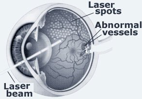 Laser treatment of diabetic retinopathy
Virectomy: In advanced PDR, the ophthalmologist may recommend a virectomy. During this microsurgical procedure, which is performed in the operating room, the blood-filled vitreous is removed and replaced with a clear solution. The ophthalmologist may wait for several months or up to a year to see if the blood clears on its own before performing a virectomy.
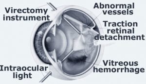 Vitrectomy Surgery
Virectomy often prevents further bleeding by removing the abnormal vessels that caused the bleeding. If the retina is detached, it can be repaired during the virectomy surgery. Surgery should usually be done early because macular distortion or traction retinal detachment will cause permanent vision loss. The longer the macular is distorted or out of place, the more serious the vision loss will be.
Vision loss is largely preventable
If you have diabetes, it is important to know that today, with improved methods of diagnosis and treatment, only a small percentage of people who develop retinopathy have serious vision problems. Early detection of diabetic retinopathy is the best protection against loss of vision.
You can significantly lower your risk of vision loss by maintaining strict control of your blood sugar and visiting your ophthalmologist regularly.
When to schedule an examination
People with diabetes should schedule examinations at least once a year. More frequent medical eye examinations may be necessary after the diagnosis of diabetic retinopathy.
Pregnant women with diabetes should schedule an appointment in the first trimester because retinopathy can progress quickly during pregnancy.
If you need to be examined for classes, it is important that your blood sugar be in consistent control for several days when you see your ophthalmologist. Glasses that work well when the blood sugar is out of control will not work well when sugar stable.
Rapid changes in blood sugar can cause fluctuating vision in both eyes even if retinopathy is not present.
You should have your eyes checked promptly if you have visual changes that:
* affect only one eye;
* last more than a few days;
* are not associated with a change in blood sugar.
When you are first diagnosed with diabetes, you should have your eyes checked:
* within five years of the diagnosis if you are 30 years old or younger;
* within a few months of the diagnosis if you are older than 30 years.